Research - (2021) Volume 11, Issue 3
Comparative flower micromorphology and anatomy in Hymenocallis spesiosa and Narcissus pseudonarcissus (Amaryllidaceae)
O. Fishchuk1* and A. Odintsova2Abstract
The structure of the flower parts of Hymenocallis speciosa (L. f. ex Salisb.) Salisb. and Narcissus pseudonarcissus L. were examined under light microscopy on permanent preparation of transverse and longitudinal sections of the flower. The work aimed to find out the features of the flower micromorphology and anatomy, the internal structure of the gynoecium in family members, which have not yet been studied in this aspect, and conduct a comparative analysis with the studied family members. The flowers of the studied species are three-merous and have the same structure, with a tubular perigonium, the inferior ovary, but differ in the structure of the corona (formed from stamens in Hymenocallis speciosa and perigonium in Narcissus pseudonarcissus). Stamens and tepals are six. Stamens attached to the perigonium tube. The most significant difference is shown by the vertical gynoecium zonality of the studied species, namely, the presence and relative height of the vertical zones in the ovary. Namely, in Hymenocallis speciosa, most of the ovary is formed by a hemisymplicate zone, there is no synascidiate zone in the ovary, two basal ovules, septal nectaries are located almost from the ovary base to the style base. In Narcissus pseudonarcissus, we found all zones of eusuncarpous gynoecium, synastidiate, symplicate and hemisymplicate, with many ovules and septal nectaries located at the top of the ovary. In both species, the asymplicate (apocarpous) zone forms a style above the opening of the nectary cavities to the outside and a stigma. The septal nectaries in Hymenocallis speciosa and Narcissus pseudonarcissus are long, non-labyrinthine, "lilioid"-type, with an apical output channel. The vascular system of the studied species has significant similarities, in particular, multibundled traces of tepals, formation of stamens traces, and dorsal vascular carpels bundles at the base of the inferior ovary, which supply the ovules and septal nectaries from paired ventral vascular carpels bundles which formed at the ovary base from vascular bundles of the ventral complex. Narcissus pseudonarcissus flower is similar to the representatives of the tribe Galantheae - Galanthus nivalis, and Leucojum vernum, studied earlier in the presence of synascidiate symplicate vertical ovary zones, numerous ovules, and aerenchyma in the flower organs.
Keywords
Hymenocallisspeciosa,Narcissuspseudonarcissus, septal nectary, vascular anatomy, inferior ovary, vertical zonality, syncarpous gynoecium.
Introduction
Modern molecular taxonomy in the phylogeny reconstruction of the families and genera of Amaryllidaceae is making great strides (Chase et al., 2016; García et al., 2019). In particular, the American botanist Alan Meerow is intensively engaged in molecular research of the family Amaryllidaceae (Meerow et al., 2006; Chase et al., 2009; García et al., 2019). According to current data, the family Amaryllidaceae includes three subfamilies and 14 tribes; among them, the most numerous is the subfamily Amaryllidoideae Burnett (Chase et al., 2016). The subfamily Amaryllidoideae includes genera Narcissus (tribe Narcisseae Lamarck & de Candolle) and Hymenocallis (tribe Hymenocallideae Small Andean), both have a unique flower structure – the corona, located inside the perigonium. The corona in Narcissusis formed by the tepaline tubular corona, and the corona in Hymenocallis is formed by the filament corona.
The genus Narcissus has about 50 species distributed from Europe to West Asia and North Africa, the genus Hymenocallis – also 50 species distributed in the southeastern United States, the Antilles, and southern Mexico to Bolivia. (Stevens, 2020). Both genera have many ornamental representatives that are connate in Ukraine and around the world. A phylogenetic study of the genus Narcissus using five markers from three genomes: ndhF and matK (chloroplast DNA), cob and atpA (mitochondrial DNA), and ITS (nuclear ribosomal DNA) was conducted by a team of Spanish scientists (Marques et al., 2010, 2017). It has been shown that the evolutionary consequences of natural hybridization between species can vary so dramatically depending on spatial, genetic, and environmental factors that different approaches are required to detect them.
Many scientists studied the representatives of these genera; in particular, M. Chwil (2006) studied the ecology of flowers and the morphology of pollen grains of selected varieties of Narcissus. The study included five cultivars of daffodils (Narcissus pseudonarcissus L. x Narcissus poëticus L.): Fire Bird, Hardy, Ivory Yellow, Pomona, and The Sun. The longevity of the flower and the flowering period of the studied Narcissusvarieties were determined, the floral elements, the anatomical structure of the ovary, nectaries, and the morphology of pollen grains were compared under a scanning electron microscope. The flowering period of 'The Sun' was the longest, whereas it was the shortest in 'Fire Bird' and 'Ivory Yellow'. The perigonium of the studied taxa was characterized by corolla outgrowths that differed from each other (Chwil, 2006).
Some members of the genus Narcissus have systematically studied the genetic diversity at both morphological and molecular levels (Liu, 2017). Twenty-four traits of the nine varieties of daffodils were described, and their differences were assessed by clustering. The results showed that nine species of daffodils could be divided into two subclusters: N. pseudonarcissus and the other - Chinese Narcissus. Morphological diversity among the five varieties of N. pseudonarcissus is higher than among the four ecotypes of Chinese narcissus (Narcissus tazetta var. Chinensis) (Liu et al., 2017; Liu et al., 2018). This group of scientists also performed biochemical analysis and investigated volatile compounds in the perigonium and corona in different varieties of N. pseudonarcissus (Liu et al., 2017; 2018). Genetic diversity and analysis of the main components based on plant, flower, and bulb traits in daffodils have been studied (Dhiman et al., 2019; Zeybekoğlu et al., 2019).
M. Grabsztunowicz and a group of researchers studied the electron transport pathways in isolated chromoplasts from N. pseudonarcissus (Grabsztunowicz et al., 2019). The study of the conservation conditions of the rare N. pseudonarcissus was carried out by A.S. Vaz et al. (Vaz et al., 2016). Many scientists are investigating the presence of alkaloids in Narcissus pseudonarcissus (Hammoda et al., 2018; Hulcová et al., 2019; Ferdausi et al., 2020; Mamun et al., 2020), the ecophysiology of seed dormancy and control of its germination was performed by R. J. Newton and colleagues (Newton et al., 2015; Newton et al., 2020).
Tanee et al. studied the karyotype of Hymenocallis littoralis (Tanee et al., 2018). The action of methanol extracts of H. littoralis on bulbs, roots, flowers, stems, and stamens was studied by scientists from Malaysia (Sundarasekar et al., 2018). Different species and cultivars of the genus Hymenocallis, growing conditions, diseases, and their care have been studied by researchers from India (Singh et al., 2017).
Despite a comprehensive study of the genera and cultivars Hymenocallis and Narcissus, the internal structure of gynoecium and septal nectaries and the anatomy of the flower have been studied very limitedly (Daumann, 1970). Therefore, our work aims to elucidate the features of the flower morphology and the internal structure of the gynoecium and to identify its vertical zonality in the species H. speciosa and N. pseudonarcissus.
Materials and Methods
Plant material was collected in the A.V. Fomin Botanical garden of the Taras Shevchenko National University of Kyiv and fixed in 70% alcohol. Five flower buds were dehydrated in t-butanol series (20%, 30%, 50%, 70 %, 100% - 2 h each, the last one - 24 h) and stored in 100% t-butanol and paraplast in the ratio 1:1. Infiltration was performed using Paraplast (Merck®) according to manufacturer's instructions and R. P. Barykina (Barykina et al., 2004). Transverse and longitudinal sections of 20 µm thickness were obtained with a manual rotary microtome (MPS-2, USSR) and stained with Safranin (Sigma-Aldrich®) and Astra Blau (Merck®). Slides were mounted in "Eukitt®" (Sigma-Aldrich®), and images were obtained with an AMSCOPE 10MP digital camera attached to an AMSCOPE T490B-10M (USA) microscope.
For the morphological analysis, measurements were made on at least 15 fresh flowers. We used the concept of gynoecium vertical zonality by W. Leinfellner (Leinfellner, 1950) to analyze the gynoecium's internal structure, which considers only the congenital fusion of the carpels. According to this concept, with the carpels' growth, the congenital multilocular synascidiate, unilocular symplicate, transitional hemisymplicate, and asymplicate (apocarpous) zones could be formed in the syncarpous gynoecium. In conditions of incomplete fusion of carpels, a hemisyncarpous gynoecium with hemisynascidiate, hemisymplicate, and asymplicate zones form only in their outer part. Later, the method was elaborated for monocots (Novikoff & Odintsova, 2008; Odintsova, 2013). The height of the zones of gynoecium was measured according to the number of cross-sections.
Results
General morphology of the flower and micromorphology of gynoecium
The flowers of H. speciosa are 24.0-24.6 cm long. The flower scape is 48-65 cm long, 1.5 cm in diameter, and 1.2 cm at the top 10-13 flowers in the inflorescence. Bracts are cone-shaped, 7 cm and 7, 2 cm long, 2.4-3 cm wide, leathery, light brown. Pedicel is 0.2 cm long, 0.4 cm in diameter. Perigonium is slightly zygomorphic, corolla-shaped, white. The leaves of a simple perigonium grew in a long flower tube, 10-10.5 cm long, 0.5 cm in diameter. Free tepals are lanceolate, 13 cm long inner tepals and 13.5 cm long outer tepals and 0.5 cm wide, spirally connected. Stamens connate to the perigonium tube. The stamens form a broad corona 2.8-3 cm high and 6 cm in diameter. The length of the outer filaments is 12.5 cm, and the length of the inner filament is 12 cm. The stamens are 0.1 cm in diameter. Anthers are wavy, 2.2 cm and 1.7 cm long, 0.1 cm wide, inner anthers are shorter than outer anthers, connected with a filament above the middle (Fig. 2 D).
The gynoecium consists of three fused carpels, each of which has two basal ovules found by micropyles down and toward the center. The pistil is somewhat zygomorphic. The ovary is obovate 0.4 cm in diameter, 0.6 cm in height (Fig. 1A). Style is apical (Fig. 2D ), placement in the center of the ovary, filamentous, s-shaped, 22.6 cm long, 0.1 cm in diameter, white at the base, above the middle is green. Stigma is dark green, capitate, trilobate. Stigma lobes are 0.1 cm long and 0.1 cm in diameter.
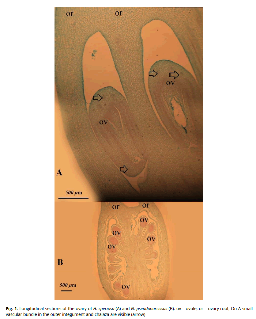
Figure 1: Longitudinal sections of the ovary of H. speciosa (А) and N. pseudonarcissus (В): ov – ovule; or – ovary roof; On A small vascular bundle in the outer integument and chalaza are visible (arrow)

Figure 2: Floral parts of H. speciosa: А – ovary wall in the median part of the carpel, dorsal vein composed of two radially located bundles and additional veins are visible; note small cells of the inner ovary epidermis in the plane of a dorsal vein; B – the central part of the ovary with three septal nectaries, paired ventral vascular bundles are visible, no stylar channel presents; C – ovary wall with septa attached, septal vascular bundle and additional bundles, D – style and anthers, triradial style channel and dorsal veins are visible (on D); dv – dorsal vein; ov – ovule; sn – septal nectary; sv – septal vein; vv – ventral vein. On A and C, small vascular bundles in the outer integument and chalaza are visible (arrow)
In H. speciosa gynoecium, we distinguish the following structural zones: a fertile multilocular zone, about 60 μm high, which corresponds to a symplicate structural zone (Fig. 6C) and a zone with a septal nectary – a hemisymplicate structural zone, the height of which is about 3340 μm (Figs. 6D, 6F). The ovary roof is 420 μm. The ovary roof has a transitional character: here, the interior style structure is formed, and the septal nectary comes out at the level of the flower tube appearance. The septal nectary appears at an altitude of 700 μm after the appearance of the ovary locules, and it is 3760 μm high (Fig. 2B, Fig. 6D-F). The asymplicate zone forms a style and a stigma (Fig. 6G).
N. pseudonarcissus flower is up to 4.5 cm long. The flower scape is 17-20 cm long and 0.4 cm in diameter. Bract is cone-shaped, folded, about 3.5 cm long, at the base in a width of 0.7 cm, above 1.2 cm, and tapers to the top. Pedicel 1, 8 cm long, 0.2 cm in diameter. Perigonium is simple, six-membered, bright yellow; tepals are different in lengths. The outer tepals are 1.8-1.9 cm long and 0.8-0.9 cm wide, and the inner tepals are 1.5-1.6 cm long and 0.7-0.8 cm wide, respectively. The perigonium is also had an elongated tube, which is formed as a result of the perigonium outgrowths and is called the corona; it is bright yellow. The flower tube is jug-shaped, 1.2 cm long, and 0.7 cm in diameter.
Stamens are connate to the perigonium tube (Fig.4 D), free at the top. The stamens of the inner circle are 1.3 cm long, and 0.1 cm in diameter, and the outer circle's stamens are 1.1 cm long and 0.1 cm in diameter. Anthers are linear; on the outer stamens, the anthers are 0.8 cm long, and on the inner 0.9 cm long and 0.1 cm in diameter, they are attached to the stamen thread below the middle of their height. The ovary is inferior, three-locular, ovoid, convex, 0.7 cm high, and 0.4 cm in diameter (Fig.1 B). There are 7-8 ovule pairs in each locule. The filamentous style has a central arrangement, 1.9 cm high and 0.1 cm in diameter. The stigma lobes are lamellar, bent, posted at three different levels, 0.1 cm long, 0.05 cm, and 0.25 cm.
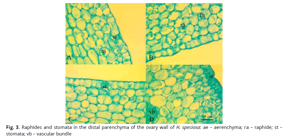
Figure 3: Raphides and stomata in the distal parenchyma of the ovary wall of H. speciosa: ае – aerenchyma; ra – raphide; st – stomata; vb – vascular bundle
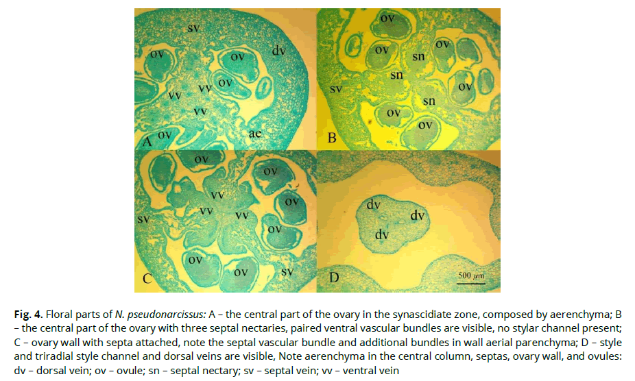
Figure 4: Floral parts of N. pseudonarcissus: A – the central part of the ovary in the synascidiate zone, composed by aerenchyma; B– the central part of the ovary with three septal nectaries, paired ventral vascular bundles are visible, no stylar channel present; C – ovary wall with septa attached, note the septal vascular bundle and additional bundles in wall aerial parenchyma; D – style and triradial style channel and dorsal veins are visible, Note aerenchyma in the central column, septas, ovary wall, and ovules: dv – dorsal vein; ov – ovule; sn – septal nectary; sv – septal vein; vv – ventral vein
The presence characterizes the gynoecium zonality in N. pseudonarcissus: at the base of the locules, there is a synacidiate structural zone – 1140 μm, three separate ovaries locules present (Fig.7 D-F). Above there is a symplicate zone, which contains ovules (Fig.7 G). This zone in the ovary is about 820 μm. Above there is the hemisymplicate zone (Fig.7 H-I) is the longest, 2080 μm high, which occupies the upper part of the locule. The septal nectary appears at an altitude of 1960 μm from the beginning of the ovary locule. The total height of the septal nectary is 2080 μm; the septal nectary opens at the flower tube base (Fig. 4 B). The asymplicate zone forms a style and a stigma (Fig.7 K-L).
Flower anatomy
In the upper part of the peduncle of H. speciosa, at the base of the flower tube, in the stamen filaments, and the ovary wall, there are idioblasts with cellular inclusion - raphides (Fig. 3). They are absent in the free tops of the tepals, connective, and the style. The stomata are present on the surface of the inferior ovary wall. The wall of the inferior ovary is formed of about 25 layers of cells.
At the peduncle base in H. speciosa there is a continuous vascular cylinder (Fig. 6 A). Slightly higher at the base of the perigonium deviate nine vascular bundles to the inner tepals and eight vascular bundles to the outer tepals and six stamens traces; all vascular bundles depart separately from the vascular cylinder. At the level of the ovary base, three massive vascular bundles are deflected - the dorsal vascular bundles and in the center remains a large number of small vascular bundles, which diverge on both sides of the septal nectary – ventral complex (Fig. 6 B). Above at the level of ovule attachment, these small bundles merge to form 6 massive vascular bundles – the ventral vascular bundles of which supplied ovules. (Fig. 6 C-E). Above the locules, the ventral vascular bundles merge with the dorsal vascular bundles to form dorsal veins that do not branch until the stigma (Fig. 6 F). The ovule traces are branches in the chalaza; its numerous branches take place in the outer integument, thickened closer to the chalazal pole, on average ten layers, the inner integument is two-layered. (Fig. 1 A, Fig. 2 A, C).
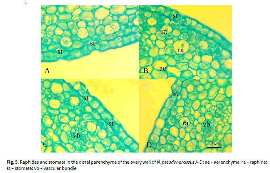
Figure 5: Raphides and stomata in the distal parenchyma of the ovary wall of N. pseudonarcissus A-D: ае – aerenchyma; ra – raphide; st – stomata; vb – vascular bundle
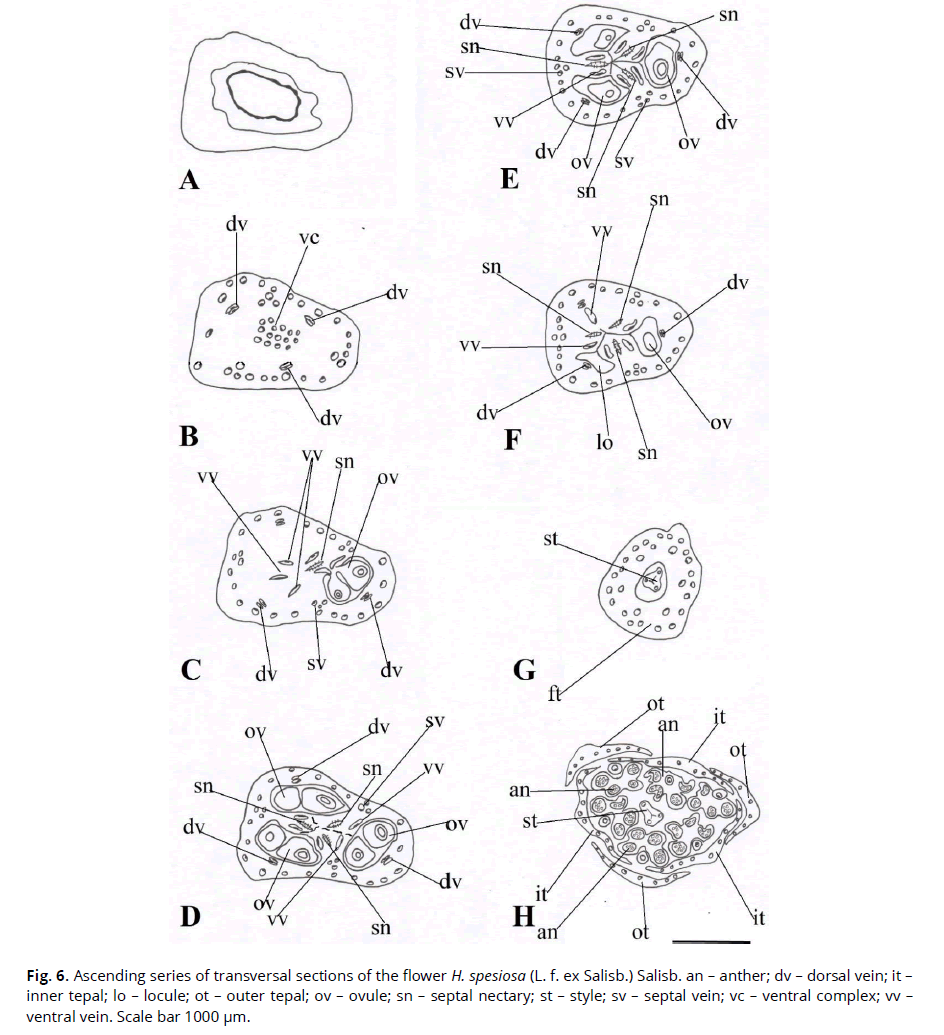
Figure 6: Ascending series of transversal sections of the flower H. spesiosa (L. f. ex Salisb.) Salisb. an – anther; dv – dorsal vein; it – inner tepal; lo – locule; ot – outer tepal; ov – ovule; sn – septal nectary; st – style; sv – septal vein; vc – ventral complex; vv – ventral vein. Scale bar 1000 μm.
General anatomy of N.pseudonarcissusis characterized by the presence of stomata in the outer epidermis of the ovary. In the upper part of the peduncle of N. pseudonarcissus, at the base of the flower tube, and in the wall of the ovary, the free tips of the tepals, there are cellular inclusions - (Fig. 5). They are absent in stamens, connectives, and style. In the ovary wall, in the ovary septa, in the central column, in filaments, in ovules, there is spongy parenchyma - aerenchyma. The wall of the inferior ovary is formed of about 20 layers of cells (Fig. 5). At the base of the peduncle of N. pseudonarcissus, there is a vascular cylinder. Even higher in the peduncle of N. pseudonarcissus, the vascular cylinder is divided into seven large vascular bundles and many small vascular bundles located around the large ones – cortical vascular bundles (Fig. 7 A-B). At the receptacle level, large vascular bundles form a vascular cylinder of the circular form, from which the dorsal vascular bundles departs (Fig. 7 C). At the locule level in the center of the ovary, a triangle is formed from small vascular bundles – the roots of the ventral complex (Fig. D-E), which turn into three crescent-shaped vascular bundles. Xylem is located outside of these bundles (Fig. 7 F). Even higher, the roots of the ventral complex disintegrate to form paired ventral vascular bundles and small blind bundles in the center of the ovary (Fig. G-H). At the level of septal nectaries, numerous small bundles are formed from the ventral bundles, which surround the nectary cavities (Fig.7 H-I). Each ovule is supplied by one vascular bundle. Above the locule, the ventral vascular bundles fuse with the dorsal ones and form a dorsal vein (Fig. 7 J-K). Traces of stamens are single-bundled (Fig.7 L).
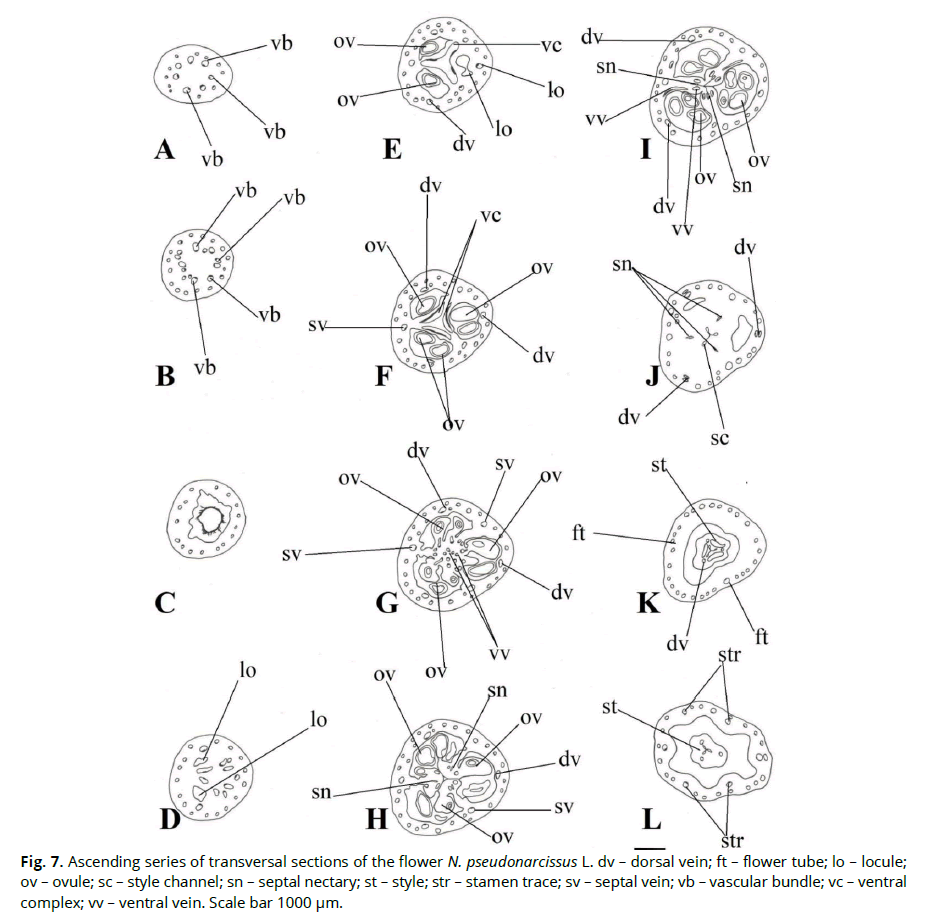
Figure 7: Ascending series of transversal sections of the flower N. pseudonarcissus L. dv – dorsal vein; ft – flower tube; lo – locule; ov – ovule; sc – style channel; sn – septal nectary; st – style; str – stamen trace; sv – septal vein; vb – vascular bundle; vc – ventral complex; vv – ventral vein. Scale bar 1000 μm.
Discussion
The genus Hymenocallis flowers are usually sessile, erect, crater-shaped, actinomorphic, white, fragrant. The stamens are always placed in a prominent funnel or rotated in a pure white corona, extended, free, straight stamens but visible to the outside. Pollen is spherical; the grains are substantial and auricular. Stigma is headed. Ovules are 2-10 in each locule. Seeds are sometimes polyembryonic (Meerow & Snijman, 1998).
The genus Narcissusis characterized by the presence of one or more upright or descending flowers. Perigonium is white or yellow, sometimes two-colored, rarely green, from actinomorphic to slightly zygomorphic crater-shaped; flower tube from inverted conical to cylindrical. Stamens in 1-2 circles, attached to the base of the flower tube, straight or inclined ascending, anthers basifix. Stigma is pronounced and trilobate. The capsule is berry-like, dehiscent; seeds are spherical, angular and flattened, sometimes with an elaiosome and black testa (Meerow & Snijman, 1998).
The corona of the genus Hymenocallis is formed by paired outgrowths of stamen filaments without vascular bundles, and the corona of the genus Narcissusis tubular, contains vascular bundles (Scotland, 2013), and is not associated with the stamens (Arber, 1937).
In cross-pollinated flowers, which are usually drooping and shallow, nectar flows freely; it is stored in the depth of the corolla in flowers adapted to specialized, ligulate or beaked animals. In most cases, the container is a perigonium tube. It can be extraordinarily elongated and erect in moth-pollinated taxa, as in the genus Hymenocallis (Vogel, 1998).
Heterostyly, namely, unequal length of style and stamen filaments in flowers of the same plant species in the genus Narcissus, promotes cross-pollination between different morphs (Barrett, 1993, 1995). It was found that in Narcissus tazetta with genetically based flower polymorphism, in different growth environments, the length of the style differed significantly, depending on local pollinators; in one case, it was hawks, in another bees and flies. Moreover, the difference in nectar concentration may be adaptive for the pollination of flowers with long and short styles (Arroyo, 1995). Thus, the large size, tubular perigonium, and nectaries in the studied flowers show their adaptation to cross-pollination by long-stemmed insects.
The morphological characteristics of the studied species were diversified in different directions on different continents, where the areas of the genera Hymenocallis and Narcissus lie.
Thus, the flowers of H. speciose are lateral, sessile, slightly zygomorphic, gathered in many flowered inflorescences. Narcissus flowers are apical, arranged singly with a leafless peduncle, drooping. However, both have inferior ovaries and fused tepals. The most significant differences are revealed in the vertical gynoecium zonality of the studied species. Thus, in H.speciose, most of the ovary is formed by a hemisymplicate zone; the ovules are paired, basal, and septal nectaries are located almost at the base of the ovary. In Narcissuspseudonarcissus,we found all zones of eusyncarpous gynoecium, synascidiate, symplicate and hemisymplicate, many ovules, and septal nectaries located in the upper part of the ovary. In both species, the asymplicate (apocarpous) zone forms a style above the opening of the nectary outlet channels on the outside.
The gynoecium structure in N. pseudonarcissus is very similar to the gynoecium structure in other family members: Amaryllidaceae - Galanthus nivalis, Leucojum vernum from the tribe Galantheae, but in these species, there are no septal nectaries (Fishchuk, Odintsova, 2020). The presence of a synascidiate zone in the ovary resembles the structure of the gynoecium N. pseudonarcissus in species of the genus Allium (subfamily Allioideae) (Shamrov, 2010).
Examining the structure of the flowers H. speciose and N.pseudonarcissus, we found that the septal nectaries are quite long, which is an adaptation to insect pollination. According to Schmid's classification (Schmid, 1985), such nectaries belong to the "lilioid" separate non-labyrinthine type. Amaryllidaceae have previously been reported to have no septal nectaries present in the upper ovary part (Daumann, 1970; Rudall, 2002).
The flower of the studied species at the anthesis stage has many parenchymal tissues in these cells of which raphide inclusions occur singly. The inferior ovary wall of the studied species at the anthesis stage is multilayered, green, that performs photosynthesis, as evidenced by the presence of stomata on the outer ovary epidermis. A flower anatomy feature of N. pseudonarcissus is a large number of aerenchyma cells in the flower organs, including the ovules, similar to what we found in Galanthus nivalis and Leucojum vernum (Fishchuk, Odintsova, 2020).
The vascular system of H.speciosaand N.pseudonarcissusis characterized by a continuous vascular cylinder in the peduncle, from which the tepal traces, stamens, and dorsal vascular bundles of the carpels depart. The ascending vascular bundles pass in the inferior ovary wall in one circle or two close circles. Tepals traces are multibundled, similar to Galanthus nivalis and Leucojum vernum (Fishchuk, Odintsova, 2020). Traces of stamens are formed at the ovary base, regardless of the tepal traces. An interesting phenomenon is the branching of the ovule trace in the chalaza, which is associated with reducing the ovule number to 2 in the locule and the large size of the ovules. The outer integument, in which goes the branching ovule traces, is multilayered. At the level of the ovary base in the center of the flower remains a large number of small vascular bundles - the ventral complex. At the level of ovary attachment, these small bundles merge to form 6 massive vascular bundles - ventral vascular carpel bundles, which supply the ovules with their branches.
The vascular system of the peduncle N. pseudonarcissus differs from H. speciosa by the presence of cortical bundles, and it has a different organization of the ventral complex. Thus, in the center of the ovary is formed a vascular triangle of small vascular bundles - the roots of the ventral complex, which are transformed into three crescent-shaped vascular bundles, in which the xylem is located outside. Ventral carpel bundles are formed from these parts of the ventral complex. The ovule trace does not branch.
Morphologists are looking for new morphological features, vascular anatomy, and flower features and study the morphogenesis of the flower-fruit system (Odintsova et al., 2017; Skrypec Odintsova 2020; Andreychuk et al., 2020; Phillips et al., 2020). It is impossible to study only a flower. At the same time, the fruit has some morphological fruit features that arise at a flower stage, for example, a double dorsal vein which testifies to the further development of loculicidal fruit opening which studying at a flower stage does not give us f clear idea of the feasibility of such a structure. Therefore, the studied
micromorphology and anatomy and flower features were analyzed in terms of fruit adaptation. In the Amaryllidoideae family, dry fruits are prevalent - loculicidal capsules, occasionally juicy capsules. Loculicidal capsules are characterized by the presence of a mechanical cell layer in the pericarpium. Thus, in cultivated Narcissus forms, the fruit is a dry capsule with woody endocarp, with U-shaped thickening of the cell walls, and the dorsal vein has V-shaped xylem (Rasmussen et al., 2006). We also found a double dorsal vein in Galanthus nivalis and Leucojum vernum (Fishchuk, Odintsova, 2020). In H. speciose and N. pseudonarcissus, the beginning of endocarp lignification is not detected at the anthesis stage, and the dorsal vein is not double.
Conclusion
Our micromorphology and anatomy study of the flowers H. speciose and N. pseudonarcissus revealed differences in the vertical zonality and gynoecium vascularization of these species. The flower of H. speciose does not contain a synascidiate zone in the ovary, the locule with two ovaries. N. pseudonarcissus flower is similar to the representatives of the Galantheae tribe - Galanthus nivalis, and Leucojum vernum in the presence of synascidiate and symplicate ovary vertical zones, numerous seeds, and aerenchyma in the flower organs. The septal nectary in Hy. speciose and N. pseudonarcissus is long, non-labyrinthine, "lilioid"- type, located almost from the ovary base in H.specioseand above mid-height N.pseudonarcissus. The vascular system of the studied species has significant similarities, in particular, multibundled traces of tepals, formation, stamens traces and dorsal vascular carpels bundles at the inferior ovary, base, supply of ovules, and septal nectaries from paired ventral carpellary bundles formed at the base of bundles of the ventral complex.
Acknowledgments
The article is made within the state theme framework: "Comparative flower and fruit morphology in Amaryllidaceae J.St.-Hil. in connection with taxonomy issues" (State registration number 0120U101743).
References
Arber, A. (1937). Studies in flower structure III. On the corona and androecium in certain Amaryllidaceae. Annals. Botany.II, 1, 293–304. Arroyo, J., & Dafni, A. (1995). Variations in habitat, season, flower traits and pollinators in dimorphic Narcissus tazetta L. (Amaryllidaceae) in Israel. New Phytologist, 129, 135–146. doi: 10.1111/j.1469-8137.1995.tb03017.x
Barrett, S.C.H., Lloyd, D.G., & Arroyo, J. (1996). Stylar Polymorphisms and the Evolution of Heterostyly in Narcissus (Amaryllidaceae). In: Lloyd D.G., Barrett S.C.H. (eds) Floral Biology. Springer, Boston, MA. doi: 10.1007/978-1-4613-1165-2_13
Barrett, S.C.H. (1993). The evolutionary biology of tristyly. In Oxford Surveys in Evolutionary Biology, 9, pp. 283–326.
Barykina, R. P., Veselova, T. D., Deviatov, A. G., Djalilova, H. H., Iljina, G. M., & Chubatova, N. V. (2004). Spravochnik po botanicheskoy mikrotehnike. Osnovyi i metodyi [Handbook of the botanical microtechniques]. Izdatelstvo Moskovskogo universiteta, Moskva (in Russian).
Chase, M. W., Christenhusz, M. J. M., Fay, M. F., Byng, J. W., Judd, W. S., Soltis, D. E., Mabberley, D. J., Sennikov, A. N., Soltis, P. S., & Stevens, P. F. (2016). The angiosperm phylogeny group. An update of the angiosperm phylogeny group classification for the orders and families of flowering plants APG IV. Botanical Journal of the Linnean Society, 181, 1–20. doi:10.1111/boj.12385
Chase, M. W., Reveal, J. L., & Fay, M. F. (2009). A subfamilial classification for the expanded asparagalean families Amaryllidaceae, Asparagaceae and Xanthorrhoeaceae. Botanical Journal of the Linnean Society, 161(2), 132–136. doi:10.1111/j.1095- 8339.2009.00999.x
Chwil, M. (2006). Ecology of flowers and morphology of pollen grains of selected Narcissus varieties (Narcissus pseudonarcissus x Narcissus poëticus). Acta Agrobotanica, 59, 107–122. doi:10.5586/aa.2006.011
Daumann, E. (1970). Das Blütennektarium der Monocotyledonen unter besonderer Berücksichtigung seiner systematischen und phylogenetischen Bedeutung. Feddes Repertorium, 80(7-8), 463–590.
Dhiman, M. R., Kumar, S., Parkash, C., Gautam, N., & Singh, R. (2019). Genetic diversity and principal component analysis based on vegetative, floral and bulbous traits in narcissus (Narcissus pseudonarcissus L.). International Journal of Chemical Studies, 7(1), 724-729
Ferdausi, A., Chang, X., Hall, A., & Jones, M. (2020). Galanthamine production in tissue culture and metabolomic study on Amaryllidaceae alkaloids in Narcissus pseudonarcissus cv. Carlton Industrial Crops and Products, 144 (112058), 1–12. doi: 10.1016/j.indcrop.2019.112058
Fishchuk, O. S., & Odintsova, A. V. (2020). Micromorphology and anatomy of the flowers of Galanthus nivalis and Leucojum vernum (Amaryllidaceae). Regulatory Mechanisms in Biosystems, 11(3), 463–468. doi:10.15421/022071
Fishchuk, O., Odintsova, A., & Sulborska, A. (2014). Gynoecium structure in Dracaena fragrans (L.) Ker Gawl., Sansevieria parva N.E. Brown and Sansevieria trifasciata Prain (Asparagaceae) with septal emphasis on the structure of the septal nectary. Acta Agrobotanica, 66(4), 55–64. doi: 10.5586/aa.2013.051
García, N., Meerow, A. W., Arroyo-Leuenberger, S., Oliveira, R. S., Dutilh, J. H., Soltis, P. S., & Judd, W. S. (2019). Generic classification of Amaryllidaceae tribe Hippeastreae. Taxon, 68(3), 425–612. doi:10.1002/tax.12062
Grabsztunowicz, M., Mulo, P., Baymann, F., Mutoh, R., Kurisu, G., Sétif, P., Beyer, P. & Krieger‐Liszkay, A. (2019). Electron transport pathways in isolated chromoplasts from Narcissus pseudonarcissus L. The Plant Journal, 99, 245–256. doi: 10.1111/tpj.14319
Hammoda, H. M., Abou-Donia, A. H., Toaima, S. M., Shawky, E., Kinoshita, E., & Takayama H. (2018). Phytochemical and biological investigation of Narcissus pseudonarcissus cultivated in Egypt. Records of Pharmaceutical and Biomedical Sciences, 2(1), 26–34. doi: 10.21608/rpbs.2018.3769.1002
Hulcová D., Maříková J., Korábečný J., Hošťálková A., Jun D., Kuneš J., Chlebek J., Opletal L., Simone A. De, Nováková L., Andrisano V., Růžička A., & Cahlíková L. (2019). Amaryllidaceae alkaloids from Narcissus pseudonarcissus L. cv. Dutch Master as potential drugs in treatment of Alzheimer's disease. Phytochemistry, 165, 112055. doi: 10.1016/j.phytochem.2019.112055
Leinfellner, W. (1950). Der Bauplan des syncarpen Gynoeceums. Österreichische Botanische Zeitschrift, 97, 3-5, 403–436.
Liu, X., Tang, D., & Shi, Y. (2018). Volatile compounds in perigonium and corona of Narcissus pseudonarcissus cultivars. Natural Product Research, 33(24), 1–4. doi:10.1080/14786419.2018.1499632
Liu, X., Tang, D., Du, H., &Shi, Y. (2018). Transcriptome sequencing and biochemical analysis of perigoniums and coronas reveal flower color formation in Narcissus pseudonarcissus. International Journal of Molecular Sciences, 19, 4006. doi:10.3390/ijms19124006
Liu, X., Zhang, X., Shi, Y., & Tang, D. (2017). Genetic diversity analysis of nine narcissus based on morphological characteristics and random amplified polymorphic DNA markers. HortScience: a publication of the American Society for Horticultural Science, 52(2), 212–220. doi: 10.21273/HORTSCI11171-16
Mamun, A. Al, Maˇríková, J., Hulcová D., Janoušek, J., Šafratová, M., Nováková, L., Kuˇcera, T., Hrabinová, M., Kuneš, J., Korábeˇcný, J., & Cahlíková, L. (2020). Amaryllidaceae alkaloids of belladine-type from Narcissus pseudonarcissus cv. Carlton as new selective inhibitors of butyrylcholinesterase. Biomolecules 10, 800. doi:10.3390/biom10050800
Marques, I., Feliner, G. N., Draper Munt, D., Martins-Loução, M., & Aguilar, J. F. (2010). Unraveling cryptic reticulate relationships and the origin of orphan hybrid disjunct populations in Narcissus. Evolution 64, 2353–2368.
Marques, I., Fuertes Aguilar, J., Martins-Louçao, M.A., Moharrek, F., & Nieto Feliner, G. (2017). A three‐genome five‐gene comprehensive phylogeny of the bulbous genus Narcissus (Amaryllidaceae) challenges current classifications and reveals multiple hybridization events. Taxon, 66(4), 832–854. doi 10.12705/664.3
Meerow, A. W., & Snijman D. A. (1998). Amaryllidaceae. In: Kubitzki, K., Huber, H., Rudall, P. J. Stevens, P. S., Studzel, T. (ed.). The families and genera of vascular plants. III. Flowering plants: Monocotyledons: Lilianae (except Orchidaceae) Springer, Berlin, рр. 83–110. doi: 10.2307/4111190 Meerow, A.W., Francisco-Ortega, J., & Schnell, R.J. (2006). Phylogenetic relationships and biogeography within the Eurasian clade of Amaryllidaceae based on plastid ndhF and nrDNA ITS sequences: lineage sorting in a reticulate area? Systematic Botany, 31 (1), 42–60. doi: 10.1600/036364406775971787. JSTOR 25064128.
Newton, R.J., Hay, F.R., Ellis, R.H. (2015). Ecophysiology of seed dormancy and the control of germination in early spring-flowering Galanthus nivalis and Narcissus pseudonarcissus (Amaryllidaceae). Botanical Journal of the Linnean Society, 177, 246–262. doi: 10.1111/boj.12240
Newton, R., Hay, F. R., & Ellis, R.H. (2020). Temporal patterns of seed germination in early spring-flowering temperate woodland geophytes are modified by warming. Annals of Botany, 125(7). doi: 10.1093/aob/mcaa025
Novikoff, A., & Odintsova, А. (2008). Some aspects of gynoecium morphology in three bromeliad species. Wulfenia, 15, 13–24. Odintsova A. (2013). Dva osnovnyx typy septalnyx nektarnykiv odnodolnyx. [Two main types of septal nectaries in monocotyledons].
Visnyk Lvivskogo universytetu. Seriya biologichna, 61, 41–50. (in Ukraine)
Odintsova, A, & Fishchuk, O. (2017). The flower morphology in three Convallariaceae species with various attractive traits. Acta Agrobotanica, 70(1), 1705–1719. doi: 10.5586/aa.1705
Phillips, H. R, Landis, J.B., & Specht C. D. (2020). Floral Fusion: The Evolution and Molecular Basis of a Developmental Innovation. Journal of Experimental Botany. doi: 10.1093/jxb/eraa125/5802329
Rasmussen, F. N., Frederiksen, S., Johansen, B., Jorgensen, L. B., Petersen, G., & Seberg, O. (2006). Fleshy Fruits in Liliiflorous Monocots.
Aliso: A Journal of Systematic and Evolutionary Botany, 22(1), 135–147.
Rudall, P. J. (2002). Homologies of inferior ovaries and septal nectaries in Monocotuledons. International Journal of Plant Sciences, 163, 261–276. doi: 10.1086/338323
Schmid, R. (1985). Functional interpretations of the morphology and anatomy of septal nectaries. Acta botanica neerlandica, 4 (1), 125– 128.
Scotland, R.W. (2013). Some observations on the homology of the daffodil corona. In Wilkin P., Mayo S.T. (eds). Early events in monocot evolution. Cambridge Universal Press, Cambridge, 297–303.
Shamrov, I. I. (2010). The peculiarities of syncarpous gynoecium formation in some monocotyledonous plants. Botanical Journal. 95(8): 1041–1070.
Singh, A., & Misra, S. (2017). Hymenocallis. Commercial Ornamental Crops: Traditional and Loose Flowers, 163–170
Skrypec, K., & Odintsova, A. (2020). Morphogenesis of fruits in Gladiolus imbricatus and Iris sibirica (Iridaceae). Ukrainian Botanical Journal, 77(3), 210–224. doi: 10.15407/ukrbotj77.03.210
Stevens P. F. (2020) Angiosperm Phylogeny. Available from: http://www.mobot.org/MOBOT/research/APweb/
Tanee, T., Sudmoon, R., Siripiyasing, P., Suwannakud, K. S., Monkheang, P., & Chaveerach, A. (2018). New karyotype Information of Hymenocallis littoralis, Amaryllidaceae. Cytologia, 83(4), 437–440. doi: 10.1508/cytologia.83.437
Vaz, A.S., Silva, D., Alves, P., Vicente, J.R., Caldas, F.B., Honrado, J.P., & Lomba, A. (2016). Evaluating population and community structure against climate and land-use determinants to improve the conservation of the rare Narcissus pseudonarcissus subsp. nobilis. Annals of the Botanical Garden of Madrid, 73(1), e027.
Vogel, S. (1998). Floral biology. In: Kubitzki, K., Huber, H., Rudall, P. J. Stevens, P. S., Studzel, T. (ed.). The families and genera of vascular plants. III. Flowering plants: Monocotyledons: Lilianae (except Orchidaceae) Springer, Berlin, рр. 34–48. doi: 10.2307/4111190
Zeybekoğlu, E., Asciogul, T. K., Ilbi H., & Ozzambak, M. E. (2019). Genetic diversity of some Daffodil (Narcissus L. spp.) Genotypes from Turkey by Using SRAP Markers. Notulae Botanicae Horti Agrobotanici Cluj-Napoca, 47(4), 1293–1298. doi: 10.15835/nbha47411664
Author Info
O. Fishchuk1* and A. Odintsova22Ivan Franko National University of Lviv, Hrushevskiy Str. 4, 79005, Lviv, Ukraine
Citation: Fishchuk, O., Odintsova, A. (2021). Comparative flower micromorphology and anatomy in Hymenocallis spesiosa and Narcissus pseudonarcissus (Amaryllidaceae). Ukrainian Journal of Ecology, 11 (1), 178-187.
Received: 27-Feb-2021 Accepted: 20-May-2021 Published: 31-May-2021, DOI: 10.15421/2021_161
Copyright: This is an open access article distributed under the terms of the Creative Commons Attribution License, which permits unrestricted use, distribution, and reproduction in any medium, provided the original work is properly cited.
